Radiology
Our full-service Radiology Department provides you with the latest diagnostic imaging technologies, including digital X-rays, 3D mammograms, high-field (.7T, 1.5T and 3T) MRIs, low-dose CT scans and more. Our board-certified radiologists are integral to providing compassionate, patient-centered care in diagnostic and therapeutic imaging procedures.
We also offer interventional radiology, minimally invasive, image-guided procedures that enable us to diagnose and treat a variety of conditions. Due to the less invasive nature of these procedures, you can benefit from a reduction in procedural risk, a decrease in post-procedural pain and a shortened recovery time compared to open surgical procedures. In the area of interventional radiology, we offer treatments utilizing radioembolization and image-guided ablation.
We are fully prepared and ready to safely welcome you, following guidelines from the Centers for Disease Control and Prevention (CDC) and public health authorities, using the best practices for hygiene, infection control and social distancing. Don’t wait to get the care you need. See how we’re keeping you safe.
Quick and Convenient Results
- In addition to imaging on the hospital campus, we offer seven community-based locations in Arlington Heights, Buffalo Grove, Kildeer, Mount Prospect, Palatine and Schaumburg
- Images are available electronically for immediate review by our expert radiologists as well as your physician
- Diagnostic testing and X-ray procedures are available 24/7 for emergency/urgent care patients
- Open .7T MRI are available at our Schaumburg location
- Same-day appointments are available with your doctor referral when immediate testing is necessary
Diagnostics Provided
Bi-plane Angiography
The bi-plane angiography unit allows the interventional radiologist to guide their catheters and needles through a small incision to your blood vessels, arteries and organs.
It provides outstanding real-time 3D X-ray images and limits your exposure to radiation.
Procedures performed in the interventional radiology suite include: ablation, angiography, angioplasty and stenting, biliary procedures, drainage, embolization and vena cava filter placement.
Bone Densitometry (DEXA)
Measures the condition and density of your bones to evaluate you for osteoporosis. The bones that are most commonly tested are the spine, hip and sometimes the forearm.
Computed Tomography (CT) Scan and Computed Tomography Angiography/Dual-source CT Scan
A non-invasive medical test that helps physicians diagnose and treat many internal conditions. At NCH, we use low-dose, multi-slice CT scanners to create detailed cross-sectional images of your internal organs, soft tissues, bones and blood vessels.
A dual-source CT scan uses two X-ray sources at right angles to each other. This doubles the speed of the test so you receive less radiation while it also creates sharper images.
Mammography (Full-Field Digital)/3D Breast Tomosynthesis
A 3D X-ray examination of the breast used to detect and diagnose breast disease; it often can detect a mass or lump before it can be felt.
Tomosynthesis images multiple layers of the breast in a 3-dimensional (3D) image. Tomosynthesis helps to reduce false positives that mammograms can give and is more accurate than a regular mammogram, particularly in screening for breast cancer in women who have dense breasts.
Magnetic Resonance Imaging (MRI) and Magnetic Resonance Angiography (MRA)
MRI and MRA are tests used to produce two- or three-dimensional images of the structures inside your body, including your blood vessels. They do not require X-ray exposure, unlike computed tomography (CT) scans or angiograms.
Nuclear Medicine SPECT and SPECT/CT Imaging
These two imaging processes use radioactive materials to help determine the cause of a medical problem based on the function of the organ, tissue or bone.
SPECT imaging utilizes a special camera and computer to create unique images that pinpoint molecular activity. This type of imaging gives us the potential to identify disease processes at their earliest stages.
SPECT/CT employs two different types of imaging scans (the traditional single photon emission computed tomography (SPECT) gamma camera images and CT images). The fusion of these image sets provides the radiologists with more precise information about how the body is functioning along with the anatomic detail of the CT scan.
PET/CT scan
This is an imaging test that uses a radioactive substance called a tracer that looks for disease in the body and is different from MRI and CT scans.
A PET/CT scan includes two parts: a PET scan and a CT scan. The PET scan demonstrates function and what’s occurring on a cellular level while the CT records anatomical X-rays, showing the same area from another perspective.
Ultrasound
Ultrasound imaging uses sound waves to produce pictures of the inside of the body. The images produced can help diagnose the causes of pain, swelling and infection in the body’s internal organs. Ultrasound may also be used to examine a baby in pregnant women and the brain and hips in infants. It’s also used to help guide biopsies, diagnose heart conditions and assess damage after a heart attack. Ultrasound is noninvasive and does not use ionizing radiation.
Automated Breast Ultrasound (ABUS)
ABUS uses a wide-field-of-view ultrasound transducer that is placed on your chest to scan the entire breast while you are lying down. This advanced system produces a 3D image that can “see through” dense breasts to reveal areas that the radiologist was not able to view with enough precision on your mammogram.
Virtual Colonoscopy (CT Colonography)
If a polyp, lesion or tumor is identified during a colonoscopy, CT colonography can be utilized to provide 2D and 3D images of the lumen of the large intestine. The radiologist uses advanced post-processing and tools for quantitative analysis of suspected polyps and other abnormalities.
Digital Radiography (X-ray) and Digital Fluoroscopy
These imaging processes involve passing a small amount of radiation to a particular part of the body to create an image of dense and soft tissues in the body. The imaging is performed utilizing digital electronic plates and a digital capturing device in place of traditional X-ray film. This allows X-ray images to be obtained almost instantly, with greater detail and reduced radiation exposure.
Shed The Shield
No more shielding means improved diagnostic accuracy and minimal radiation
NCH is no longer use lead aprons or gonad shields when performing X-rays, CT scans or fluoroscopy on pediatric and adult patients.* Professional radiology associations, including the American College of Radiology and the American Association of Physicists in Medicine, support research that shows radiation levels used in today’s X-ray exams pose no risk to reproductive organs. In fact, patient shields can actually prevent doctors from seeing organs and parts of the body that are being examined with an imaging test. Inconclusive test results can lead to repeat exams and more radiation exposure.
Our state-of-the-art equipment uses industry leading radiation-dose reduction technology. Also, our radiology experts perform quality measures to ensure all of our diagnostic imaging technology uses the lowest levels of radiation possible for our patients’ exams.
The no-shielding policy is also endorsed by the Image Gently Alliance, whose mission is, through advocacy, to improve safe and effective imaging care of children worldwide.
*While not preferred, if you insist that we use a shield, we will honor your request if it is possible to do so without compromising the exam you are having.
Online Scheduling
You can now schedule the following services in MyChart without an order:
- Mammography screening
Additionally, you can schedule or request the following appointments if you have an order in MyChart from your provider:
- Bone density (DEXA) exam
- Diagnostic imaging (CT, PET/CT, ultrasound, or X-ray)
Log in or sign up now to enjoy the benefits of MyChart.
Expert Staff
At NCH, we are extremely proud that all of our technologists have met the standards set by many national accrediting organizations, including the American Registry of Radiologic Technologists (ARRT) and the American Registry for Diagnostic Medical Sonography (ARDMS) and remain in good standing with continued training and education. Their technical expertise is a primary reason why our patients receive high-quality imaging. The accuracy of the interpretation and diagnosis made by our radiologists is dependent on the technologists creating quality images. Our technologists embrace their role as compassionate front-line caregivers, demonstrating commitment to advancing quality health care through the power of teamwork and ensuring every patient receives exceptional care.
Radiology Treatment Locations

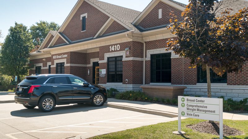
1410 N. Arlington Heights Road
Suite 100
Arlington Heights, IL 60004
847-618-3700
X.X mi


880 W. Central Road
Floor 3
Arlington Heights, IL 60005
847-618-3700
X.X mi


800 W. Central Road
Arlington Heights, IL 60005
847-618-1000
X.X mi

519 S. Roselle Road
Schaumburg, IL 60193
847-618-7850
X.X mi